Compact Bone Diagram Microscope / 1 / Saw microtome preparation procedure 1.. Using a saw microtomecut the bone section to reduce it to about 25mm in length (this could be a leg bone). Themetaphysis, which is the point between the shaft and epiphysis, is often thepoint of growth during development. Compact bones make up 80 percent of the human skeleton; 0 0000 a shoutout is a way of letting people know of a. See full list on microscopemaster.com
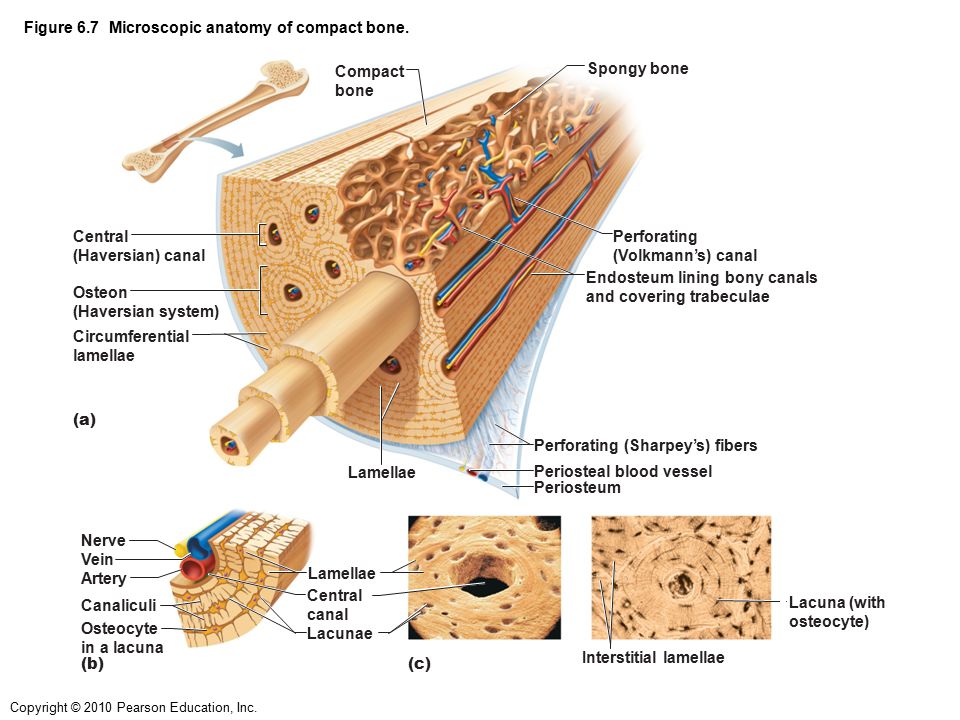
See full list on microscopemaster.com The compact bone is composed of calcified extracellular material the bone matrix and 3 major cell types which are osteoblast which ssynthesize and secrete the organic components of bone matrix which include type 1 collagen fibers proteoglycans and several glycoproteins such as ostepnectin. Each central canal contains blood vessels and nerve fibers surrounded by loose connective tissue. The osteon consists of a central canal called the osteonic (haversian) canal, which is surrounded by concentric rings (lamellae) of matrix. Osteoblasts make and secrete collagen, giving bone a measure of elasticity.
Pin By Danielle Papas On Nursing Anatomy Physiology Compact Bone Nursing Anatomy And Physiology Connective Tissues from i.pinimg.com The inner layer consists mainly of osteoblasts, cells that are constantly renewed in the bone. They serve to provide support and stability andinclude such bones as the carpal and tarsal bones. Untramicrotome with a diamond knife 8. They are also important inthat they help rebuild bones in the event that they break. See full list on microscopemaster.com *thismethod does not require significant preparation of the bone Cortical bone is compact bone, while cancellous bone is trabecular and spongy bone. These are importantfeatures of the bone in that they hold vessels through which blood andlymph are circulated.
The threemain types of cells that make up bone tissue include:
The osteon consists of a central canal called the osteonic (haversian) canal, which is surrounded by concentric rings (lamellae) of matrix. Each central canal contains blood vessels and nerve fibers surrounded by loose connective tissue. Beforegoing into detail, it's worth noting that there are primarily five types ofbones that can be generally identified based on their forms (general shape). The lacunas can also beviewed as connected to each other through what seems like very thin lines.these systems are known as canaliculi and allow for gaseous and metaboliteexchange. Using clear epox glue, bind the section to the microscope glass slide 7. Wash the bone sample using saccharose solution overnight 3. Untramicrotome with a diamond knife 8. Nov 13, 2015 · the osteons' center is a hollow canal that acts as a central passageway for blood vessels and nerves. Fix the sample in glutaraldehyde for about 2 hours 2. They serve to provide support and stability andinclude such bones as the carpal and tarsal bones. *thismethod does not require significant preparation of the bone This ensures that the cells are continually nourished andremain healthy. These processesinvolve the cell (osteoblasts) accumulating at a give spot to form osteoid(flexible material) that hardens when material (minerals) are added to it.
Wash the bone sample using saccharose solution overnight 3. Compact bones make up 80 percent of the human skeleton; Examples of flat bonesinclude ribs, scapulae and skull b. See full list on microscopemaster.com The threemain types of cells that make up bone tissue include:
Compact Bone Under Microscope Diagram Quizlet from o.quizlet.com What is the compact bone used for? The small, dark spots (lacunas) that can also be seen containosteoblast cells that form matrix and collagen fibers. The threemain types of cells that make up bone tissue include: Begin by identifying the concentric rings of lamellar bone that surround a haversian canal. If you look at compact bone under the microscope, you will observe a highly organized arrangement of concentric circles that look like tree trunks. They are also important inthat they help rebuild bones in the event that they break. Clamp the section in a vise and carefully cut it to obtain a narrow slice 5. The lacunas can also beviewed as connected to each other through what seems like very thin lines.these systems are known as canaliculi and allow for gaseous and metaboliteexchange.
Each central canal contains blood vessels and nerve fibers surrounded by loose connective tissue.
See full list on microscopemaster.com These pores serve to hold not only some marrow, butalso nerves and vessels that transport blood to the cells deliveringnourishment and gas exchange. They are also important inthat they help rebuild bones in the event that they break. Clean the bone using some warm water 3. Preparation to view a bone tissue under the microscope, the bone sample has to be carefully prepared in order to produce a specimen that will provide the best possible results. Bonetissueis one of the main components of the skeletal system (other componentsinclude bone marrow/marrow cavity, collagen fibers etc). The small, dark spots (lacunas) that can also be seen containosteoblast cells that form matrix and collagen fibers. *thismethod does not require significant preparation of the bone Under the microscope, bone can be divided into two types compact bone forms the outer 'shell' of bone. As such, they may be described as. The osteon consists of a central canal called the osteonic (haversian) canal, which is surrounded by concentric rings (lamellae) of matrix. Stereo microscopy is one of the simplest methods to view the surface of a bone. They are derived from osteoprogenitor cells and areresponsible for building new bones as one grows.
Using a ultramicrotome that is equipped with a diamond knife, cut the section again to obtain ultrathin sections 8. See full list on microscopemaster.com As mentioned, conduits referred to ashaversian canals are at the center of these layers. These processesinvolve the cell (osteoblasts) accumulating at a give spot to form osteoid(flexible material) that hardens when material (minerals) are added to it. The osteon consists of a central canal called the osteonic (haversian) canal, which is surrounded by concentric rings (lamellae) of matrix.
Microscopic Anatomy Of Compact Bone Anatomy Drawing Diagram from slideplayer.com Beforegoing into detail, it's worth noting that there are primarily five types ofbones that can be generally identified based on their forms (general shape). Therefore, osteocytes remain embedded inside the bone as new bonecontinues to form. Fix the sample in glutaraldehyde for about 2 hours 2. If you look at compact bone under the microscope, you will observe a highly organized arrangement of concentric circles that look like tree trunks. Each group of concentric circles (each "tree") makes up the microscopic structural unit of compact bone called an osteon (this is also called a haversian The osteon consists of a central canal called the osteonic (haversian) canal, which is surrounded by concentric rings (lamellae) of matrix. Stain the section using toluidine blue 7. Compact bones make up 80 percent of the human skeleton;
See full list on microscopemaster.com
Learn vocabulary, terms, and more with flashcards, games, and other study tools. How is a compact bone different from spongy bone? Compact bone, also called cortical bone, is the hard, stiff, smooth, thin, white bone tissue that surrounds all bones in the human body. As mentioned, conduits referred to ashaversian canals are at the center of these layers. *thismethod does not require significant preparation of the bone Using clear epox glue, bind the section to the microscope glass slide 7. Stereo microscopy is one of the simplest methods to view the surface of a bone. Stain the section using toluidine blue 7. What is the microscopic anatomy of a compact bone? They are derived from osteoprogenitor cells and areresponsible for building new bones as one grows. Preparation to view a bone tissue under the microscope, the bone sample has to be carefully prepared in order to produce a specimen that will provide the best possible results. Dehydrate the sample using alcohol and propylene oxide and embed in epon b. The compact bone is composed of calcified extracellular material the bone matrix and 3 major cell types which are osteoblast which ssynthesize and secrete the organic components of bone matrix which include type 1 collagen fibers proteoglycans and several glycoproteins such as ostepnectin.
Cut the section to dimensions of about 5mm by 5mm chip 6 compact bone diagram. Cortical bone is compact bone, while cancellous bone is trabecular and spongy bone.


0 Komentar